Understanding transcriptomics: A practical guide to RNA-sequencing approaches Introduction When people think about the genome, DNA is usually the first thing that comes to mind. But for genetic i...
Phân tích dữ liệu giải trình tự toàn bộ hệ gen vi sinh vật (Microbial Whole Genome Sequencing) Trong bộ gen của mỗi vi sinh vật, hàng nghìn biến thể di truyền – từ những thay đổi đơn nucle...
Giải trình tự trong nghiên cứu vi sinh (Metagenomics, Whole Genome Sequencing, Targeted Sequencing) và các ứng dụng trong lâm sàng Trong bối cảnh dịch bệnh truyền nhiễm ngày càng trở nên phức tạp và khó kiểm soát, những v�...
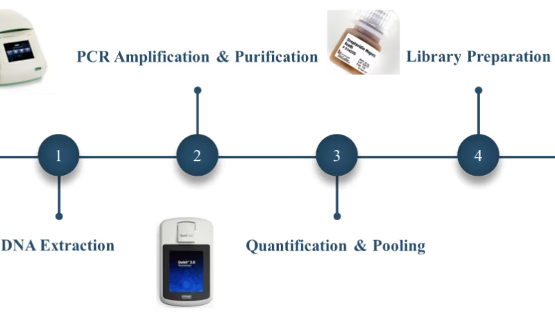
A Beginner’s Guide to Whole Genome Sequencing – Part 2: Bioinformatic analysis of standard package Continuing from our previous blog, we will dive deeper into bioinformatics analysis offered by Novogene in this part....
A Beginner’s Guide to Whole Genome Sequencing – Part 1: Introduction to WGS workflow 1. Introduction Whole Genome Sequencing (WGS) gives researchers and clinicians the most complete view o...
Kickstart Your Whole Exome Sequencing Project with Novogene 1. Introduction Whole exome sequencing, which targets the protein-coding regions of approximately 20,000 genes, is pa...
Whole Genome Sequencing vs Whole Exome Sequencing: Which Genetic Sequencing Method is Right for Your Research? 1. Introduction 1.1. Whole Genome Sequencing (WGS) Whole genome sequencing (WGS) refers to the sequencing of an en...
A Beginner’s Guide to Microbial Shotgun Metagenomic Sequencing If you’re considering between 16S/18S/ITS Amplicon Metagenomic Sequencing and need more instructions for choosing the...
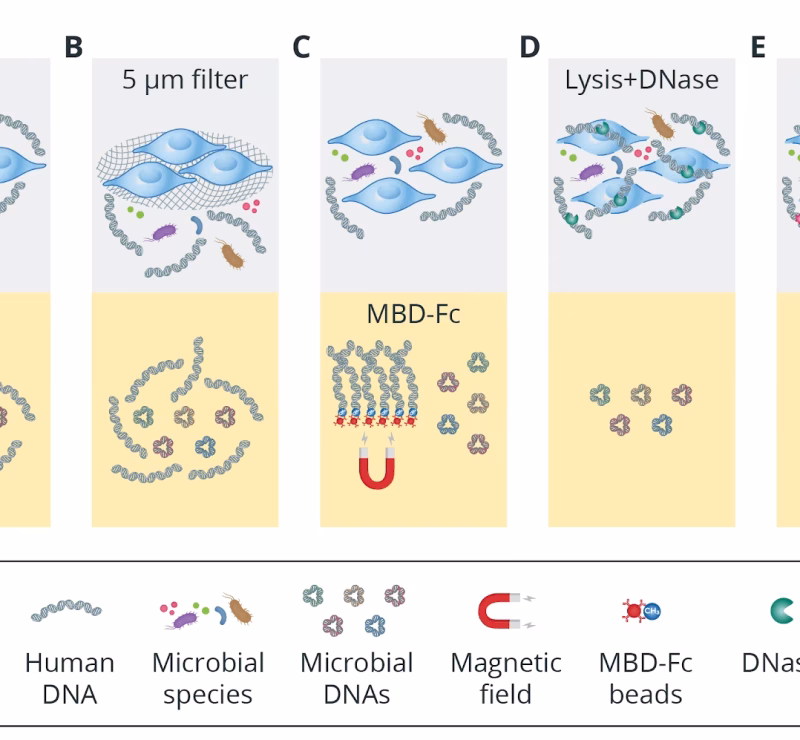
Metagenomics: Host DNA Removal or Not? 1. Introduction Over the past decade, a dramatic reduction in DNA sequencing costs has propelled shotgun metagenomic...
Amplicon vs Shotgun Metagenomic Sequencing: Choosing the right approach for Microbiome research The culture-free study of microbial community composition has been revolutionized by the application of high-throughput,...
A Beginner’s Guide to 16S/18S/ITS Amplicon Metagenomic Sequencing 1. Introduction 16S/18S/ITS amplicon metagenomic sequencing is designated to sequence the target genes of 16S ribosom...
Two platforms, one powerful spatial biology toolkit: When and how researchers are using Visium and Xenium We recently discussed the key features of our Visium Spatial Gene Expression and Xenium In Situ spatial transcriptomics...