

Optimizing your spatial transcriptomics research with Visium HD and Xenium In Situ
Novel spatial transcriptomics tools blend traditional imaging with molecular analysis to give you a more complete picture of your biology of interest. Whether you are a neuroscientist untangling the complexity of the nervous system, a translational researcher better resolving the tumor microenvironment in cancer, or trying to answer any of a myriad of other questions, spatial insights open new doors for your research projects. Given the promise of these methods and their increasing adoption, the question has morphed from, “Why should I do spatial biology?” into, “What do I want to achieve with spatial biology—and how should I do it?”
To equip you to ask the questions you want to ask, 10x Genomics have developed two complementary spatial transcriptomics methods: NGS-based whole transcriptome Visium Spatial Gene Expression (which includes “standard definition” Visium, Visium v2, and latest Visium HD) and targeted imaging-based Xenium In Situ. The question of which one is the best fit for your current applications—or if they should be used together—depends on your research project, your goals, and your practical considerations.
Technical specs
| Visium HD | Xenium In Situ | |
| Methodology | Sequenced-based spatial transcriptomics | Imaging-based spatial transcriptomics |
| Data acquisition | NGS | High-resolution microscopy |
| Analyzable area | 6.5 x 6.5 mm per tissue section (2 per Visium slide) | 22.5 x 10.5 mm per slide (2 slides per run) |
Plexity
| Visium HD | Xenium In Situ | |
| Number of RNA targets | Whole transcriptomics (≈20,000 transcripts) | Up to 480 targets (5,000 plex panel coming in Q2 2024) |
| Customization options | Custom probe spike-in for custom and exogenous genes | Up to 100 custom genes added to pre-designed panelsStandalone custom 480-plex panelsAdvanced custom probe for exogenous genes and gene isoforms |
Sensitivity
| Visium HD | Xenium In Situ | |
| Per-gene sensitivity | Gene dependent | Gene dependent but generally greater than Visium HD |
| Gene detection | Whole transcriptome | Up to 480 transcript species (genes, isoforms, etc) with 5,00-plex panel coming in 2024 |
| Transcript abundance (total transcripts per analyzed area) | Tissue dependent but generally greater than Xenium | Tissue and panel dependent |
Resolution
| Visium HD | Xenium In Situ | |
| Resolution | Single cell scale (data output at 2 x 2 μm and multiple bin sizes; 8 x 8 μm bin is a recommended starting point for analysis) | Subcellular (transcripts localized within single cells with ≈ 30 nm precision) |
| Cell segmentation | Morphology-based binning requires third-party tools | Multimodal cell segmentation performed during instrument run |
Multiomics
| Visium HD | Xenium In Situ | |
| Additional assays on same section | Pre-run: Morphology (H&E) or IF | Post-run: Morphology (H&E), iF, and Visium/Visium HD |
| High-plex protein multiomics | Release to be determined | ≤ 30 targets (coming 2024) |
Tissue fixation, organism, and tissue format
| Visium HD | Xenium In Situ | |
| Sample compatibility | FFPEFresh frozen (FF) and fixed frozen Demonstrated Protocol coming soon | Fresh frozen and FFPE |
| Organisms | Human and mouse, with non-human primate and rat possible depending on customer support guidance | Human and mouse, with other species available through Advanced Custom Design |
| Tissue format | Pre-sectioned/archived slidesTissue block (sectioned onto standard glass slides) | Tissue block (must be pre-sectioned onto Xenium slides) |
Number of samples, tissue size, and time constraints
For any technology, more samples take more time. There are, however, two major nuances to this general rule.
The first is that the extra time required does not necessarily scale linearly with each additional sample. The second is that tissue samples can be arranged on the Capture Area (Visium HD) or imageable area (Xenium) in a variety of ways—a single slide can contain one large sample, several medium samples, or many small samples

Figure: Example of tissue placement configurations on Visium HD and Xenium slides.
As an NGS-based technology, the output of each Visium HD run is a sequencing library. This allows you to batch a large number of samples in a single NGS run. Thus, Visium allows you to scale up by batching sequencing libraries from large numbers of samples. The Visium HD workflow itself requires roughly 8 hours of hands-on time over 2–3 days, and can be parallelized depending on the availability of samples, personnel, thermal cyclers, and CytAssist instruments.
Xenium is an imaging-based technology, and the most time-consuming part of any single cell spatial imaging experiment is the imaging. Tissue analysis time and throughput is a function of both total tissue area analyzed and assay plexity. For a total of 4 cm2 of tissue using a 480-plex panel with multimodal cell segmentation, we estimate 6 hours of hands-on time and 2 to 3 days of preparation for pre-image acquisition steps, which can be parallelized depending on the availability of samples, personnel, and thermal elements. For these parameters, image acquisition and analysis can take up to 3 days, though you have the option to reduce run time by choosing to analyze subsection(s) of your tissue.
We recommend you estimate each tissue sample’s size, use that to determine the total number of slides you would need to run on each technology, and leverage that to determine a rough total analysis time.
Data analysis methods
Once sequencing of your Visium libraries is completed, the data is run through the Space Ranger analysis pipeline. These outputs (which include, but are not limited to, file formats such as feature barcode matrix (h5) and tissue positions (parquet)) are then visualized with the Loupe Browser, which allows for differential analysis and reclustering of Visium clusters
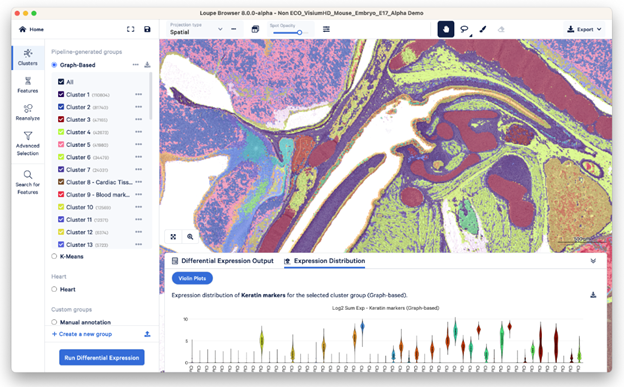
Figure: Example visualization of Visium HD data in the Loupe Browser. Visualization depicted is expression distribution of transcripts in a mouse embryo at E17.
Xenium data is analyzed onboard the instrument in parallel with the image acquisition and analysis steps, resulting in interpretable data that’s ready right after your run is done in formats like ome.tiff (images), .parquet, .zarr, and .csv (transcript). These outputs are then visualized on Xenium Explorer

Figure: Example visualization of Xenium data in Xenium Explorer. Visualization depicted is FFPE human lung cancer tissue analyzed with the Xenium Human Multi-Tissue and Cancer panel.
Both Xenium and Visium HD feature integrated and intuitive data visualization and analysis pipelines, file outputs in a variety of open formats, and easy compatibility with third-party analysis tools.
Final conclusions and taking your next steps into spatial
These technologies offer unprecedented clarity, depth, and breadth in tissue visualization. Visium HD Spatial Gene Expression enables whole transcriptome analysis with spatial context through sequencing-based spatial transcriptomics, ideal for discovering new targets or characterizing tissues broadly. For precision insights, Xenium In Situ provides nanometer-scale resolution of up to 5,000 genes within individual cells using imaging-based spatial transcriptomics with padlock probes.
Visium HD and Xenium are complementary, not alternatives. For instance, Visium HD can map gene expression in tissue, and Xenium can then provide detailed analysis of specific genes. This is exactly what Dr. Ayako Suzuki did in her latest Nature Communications publication, in which she first used Visium v2 to determine where in lung cancer tumors gene expression changes were occurring, then zoomed in with Xenium to pinpoint specific cells and cell types associated with alveolar collapse in a subtype of tumors.
Source: Optimizing your spatial transcriptomics research with Visium HD and Xenium Prime 5K
------------
GENESMART CO., LTD | Phân phối ủy quyền 10X Genomics, Altona, Biosigma, Hamilton, IT-IS (Novacyt), Norgen Biotek, Rainin tại Việt Nam.