Dive into the intricate world of tissue analysis with Visium CytAssist Gene and Protein Expression. This cutting-edge technology allows researchers to unlock a treasure trove of insights by simultaneously capturing gene expression and protein levels from a single tissue section. Say goodbye to biases and hello to a seamless workflow that integrates multiple streams of information, even from previously stained samples. With Visium’s high-resolution spatial capture technology and intuitive software, researchers can visualize gene and protein expression like never before. From understanding cellular dynamics to deciphering disease mechanisms, Visium empowers researchers across diverse fields to embark on a journey of discovery, uncovering the secrets of tissue biology and disease pathology with style and precision.
Visium CytAssist Gene and Protein Expression
Gaining a holistic understanding of tissue morphology is paramount, and nothing exemplifies this more than the simultaneous analysis of gene expression and protein levels from a single tissue section. By combining these analyses, biases introduced by separate assessments or disparate techniques like flow cytometry are eliminated.
Visium CytAssist Gene and Protein Expression revolutionizes tissue analysis by enabling the capture of barcoded antibody oligo tags and unbiased whole transcriptome gene expression alongside histological examination, all in one experiment from a single tissue section. This innovative technology offers a myriad of insights from precious samples, including previously H&E-stained and stored FFPE sections on coverslipped slides, through an integrated morphology-first workflow. Notably, this methodology allows for the identification of regions of interest post-data acquisition, ensuring that no crucial biological findings escape notice.

Figure 1. Morphology, RNA, and protein expression in one experiment, with a single tissue section. An FFPE-preserved human tonsil section was H&E stained, imaged, and then processed through the Visium CytAssist Gene and Protein Expression workflow. Shown are image overlays containing data from UMI counts of RNA, via whole transcriptome analysis, and protein. Combined analysis of morphology, RNA, and protein expression revealed canonical structural components of the human tonsil, which are labeled above.
Visium spatial capture technology
The Visium CytAssist Spatial Gene and Protein Expression assay is designed to analyze mRNA and protein in human tissue sections derived from formalin-fixed and paraffin-embedded (FFPE) tissue samples. It uses a combination of oligo-tagged antibodies (a readily available and validated 35-plex antibody panel of extracellular and intracellular immune markers and controls) and probes targeting the whole transcriptome. Each Visium CytAssist Spatial Gene Expression Slide contains capture areas with barcoded spots that include oligonucleotides necessary to capture both gene expression probes and antibody tags.


Figure 2. Visium Gene and Protein Expression slide architecture. Visium HD Spatial Gene Expression slides contain two Capture Areas with a continuous lawn of oligonucleotides arrayed in millions of 2 x 2 µm barcoded squares without gaps, achieving single cell–scale spatial resolution.
Visium Gene and Protein Expression workflow
FFPE tissue sections are placed onto standard glass slides. If needed, slides are deparaffinized and imaged with H&E or IF as in a typical FFPE workflow, then de-crosslinked and hybridized to probe pairs, followed by incubation with the validated antibody panel. The Visium CytAssist then facilitates the transfer of ligation products and antibody oligo tags from the glass slide to the capture area on a Visium CytAssist Spatial Gene Expression Slide. The captured probes and antibody oligo tags are extended to incorporate complements of the spatial barcodes, and sequencing libraries are prepared. These libraries are then sequenced, and data visualized using Space Ranger and Loupe Browser, our user-friendly data analysis and visualization software. This software allows determination of which genes and proteins are expressed, where they are expressed, and in what relative quantity. The Loupe Browser enables direct comparison of gene expression and antibody binding with H&E or immunofluorescence staining patterns.

Figure 3. Streamline experimentation with a ready-to-use, robust workflow for whole tissue section analysis of gene and protein expression.
Applications
The Visium CytAssist by 10x Genomics isn’t just a tool; it’s a gateway to discovery. With its ability to integrate spatial and molecular data, it empowers researchers across various disciplines:
- Spatially Resolve Gene Expression: Visualize and quantify gene activity within intact tissue sections to understand cell organization and function.
- Simultaneously Profile Gene and Protein Expression: Correlate mRNA data with protein abundance and localization for a comprehensive view of cellular mechanisms.
- Advance Developmental Biology: Map gene and protein expression during tissue and organ development to gain insights into growth and differentiation.
- Enhance Cancer Research: Characterize the tumor microenvironment to identify therapeutic targets and understand treatment resistance.
- Improve Neuroscience Studies: Provide high-resolution spatial data to explore brain function and neurological diseases.
- Boost Immunology Research: Profile immune cells within tissues to study immune responses and develop better immunotherapies.
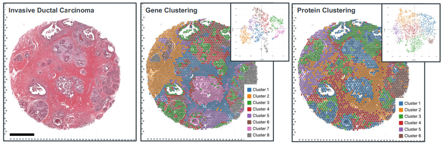
Figure 4. H&E-stained human breast tissue (DV200 = 56%) containing poorly differentiated (grade III) invasive ductal carcinoma (IDC) on Visium slide (left). Unbiased clustering of mRNA transcripts (middle) and protein (right) from the whole-transcriptome RNA panel and immunology antibody panel, respectively, superimposed on the H&E image. Both clusters demonstrated similar patterns delineating the tumor and stromal regions. Representative tSNE plots for each spatial clustering (inset). Scale bar = 1.0 mm
In the hands of researchers like Cedric R. Uytingco and Jennifer Chew, the Visium Spatial Gene and Protein Expression platform has yielded groundbreaking insights into disease progression, predictive biomarkers, drug response and resistance, and therapeutic development. It’s a testament to the power of spatially resolved, multiomic approaches in advancing our understanding of clinical tissue samples.
Source: See tissue complexity in a new light with simultaneous gene and protein spatial profiling
------------
GENESMART CO., LTD | Phân phối ủy quyền 10X Genomics, Altona, Biosigma, Hamilton, IT-IS (Novacyt), Norgen Biotek, Rainin tại Việt Nam.